Source: Nuhsbaum
What is darkfield and brightfield microscopy? It sounds like something out of Star Wars! But, unfortunately neither gives us special powers. That doesn’t mean that darkfield and brightfield microscopy aren’t still fascinating.
Not a Superpower — Super Powerful
Darkfield and brightfield microscopy are contrast-enhancement techniques in light microscopy. In simple terms, they are techniques that allow us to better see our specimens. Brightfield microscopy is the conventional technique. It is suitable for observing the natural colors of a specimen. It can also observe stained samples. The specimen appears darker on a bright background. Darkfield microscopy shows just the opposite. The specimen shows up bright on a dark background.
Darkfield Microscopy
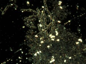
Bacteria in darkfield microscopy. Source: Wikimedia Commons
Both light and electron microscopy use darkfield microscopy methods. It excludes un-scattered light from the image to produce high-contrast images. As a result, the field around the specimen is dark because there is no specimen to scatter the light beam. Yes, you read that right! We can both illuminate the subject and darken the background… without actually shining light on the sample!
Darkfield is a simple, yet effective microscopy technique. It is well-suited for live biological samples and industrial samples. Some examples include looking at cells, single-celled organisms, starch grains, crystals, polymers, and films.
However, one limitation of darkfield is the low light levels seen in the final image. Since darkfield is caused by literally inserting a dark ring to block direct light, the light source must be at maximum intensity to get a darkfield image seen through the oculars. Looking at a black screen? Camera settings, like exposure time and gain, can increase to brighten the digital image on the monitor..
Brightfield Microscopy
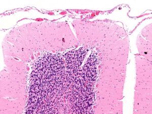
Intermediate magnification micrograph of the cerebellar cortex. H&E stain. Source: Wikimedia Commons.
Brightfield microscopy is the simplest of all the optical microscopy lighting techniques. To get better contrast in the image on dense areas of the sample, simply increase the transmitted light intensity. Then dial in the aperture on the condenser either up or down until the image looks good.
In a range of techniques for illuminating samples in light microscopes, brightfield and its simplicity makes it a popular technique. The typical appearance of a brightfield image is a dark, true color sample on a bright, white background, hence the name.
However, there are limitations. Given the nature of most biological samples, there is very low contrast and the image can look a bit whitewashed. Moreover, the limit to brightfield microscopy is around 1300X, which may not be suitable for some cases. For more contrast, try dialing down the aperture. Otherwise, phase contrast is another brightfield technique that can help.
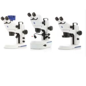
Microscope Models
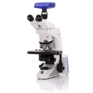
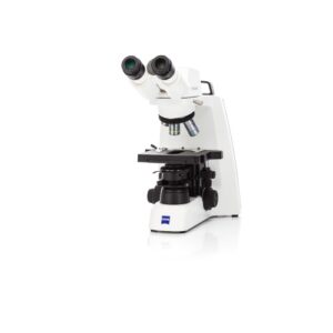
The ZEISS Primostar 3 and ZEISS Axiolab 5 compound microscopes are perfect for brightfield microscopy. Equipped with a mechanical stage to accommodate two specimen slides and labelled Abbe condenser, the Primostar and Axiolab can be configured with rotatable binocular tubes for shared viewing and easy storage. An integrated camera or trinocular tube with camera can be added so you can go digital.